Why so many people with CKD stay exhausted despite iron tablets, what the research shows about the gut, and what I look at in clinic.
If you have chronic kidney disease and you are exhausted all the time, there is a good chance anaemia is part of the story. And there is a good chance someone has already handed you an iron tablet.
Sometimes that works. Often it doesn’t. I see patients with low haemoglobin and perfectly normal iron studies, told they are “not anaemic” while they can barely get through the day. I see others who have taken iron for months, felt worse, and watched their numbers barely move.
This article digs into one piece of the puzzle that rarely gets discussed in clinic: the gut. The gut is where the body produces most of its uraemic toxins, where iron is absorbed (or blocked), and where many well-meaning iron supplements end up feeding the wrong bacteria.
As always, I’ll show you what the research says, where it’s solid, and where I’m going on clinical experience rather than settled evidence.
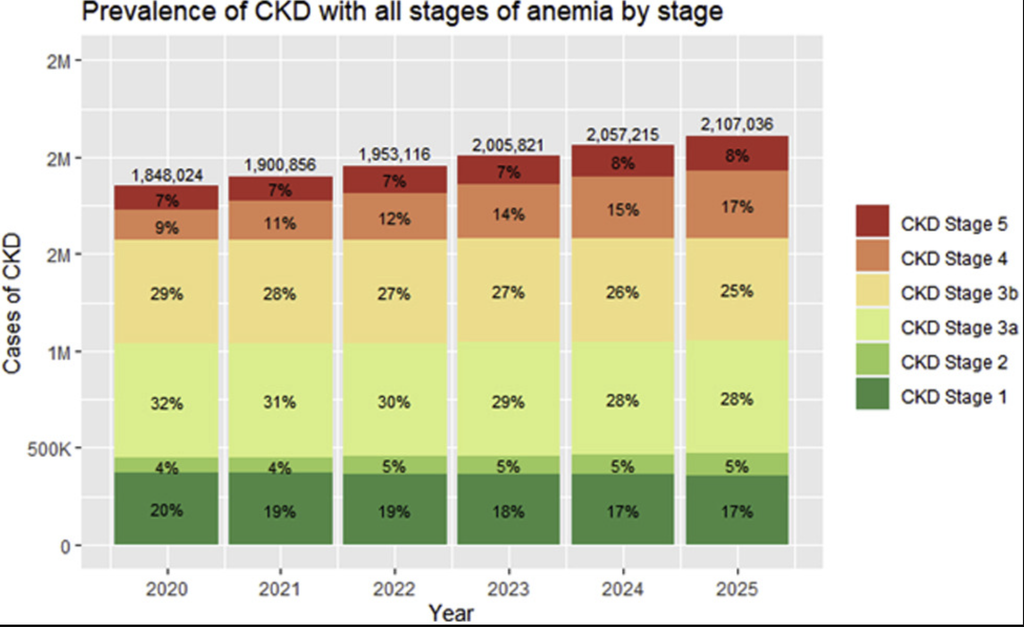
In the KNOW-CKD cohort of 2,198 non-dialysis patients, using the same haemoglobin cut-offs KDIGO now uses, anaemia affected 32.8% of people at stage 3a, 46.6% at 3b, 78.9% at stage 4 and 96.5% at stage 5 (Ryu et al., 2017). US population data using a similar definition found stage 5 prevalence closer to 53% (Stauffer & Fan, 2014).
It matters for more than energy. In a US cohort of 22,720 non-dialysis CKD patients, those with anaemia had double the rate of heart failure hospitalisation (1.6 versus 0.8 per 100 patient-years) and progressed to the next CKD stage considerably faster (43.5 versus 27.5 per 100 patient-years) (Wittbrodt et al., 2022). Those are associations, not proof of cause. But it is hard to heal a scarred, oxygen-starved kidney when the blood carrying the oxygen is thin.
And here is the part that frustrates me most: fewer than 2% of the anaemic patients in that cohort were receiving an erythropoiesis-stimulating agent (ESA). Anaemia in CKD is common, consequential, and under-treated.
In January 2026, KDIGO published its first full update of the CKD anaemia guideline since 2012. It’s better than I expected.
Anaemia is defined by haemoglobin alone: below 13 g/dL in men and below 12 g/dL in women. Normal iron studies do not mean you are not anaemic.
Screening is scheduled by stage: at least yearly at stage 3, twice yearly at stage 4 and every three months at stage 5.
The screening panel is decent: full blood count, reticulocytes, ferritin and transferrin saturation (TSAT).
Finding the cause is the next essential step. If the usual work-up doesn’t explain it, the guideline lists further tests, including B12, folate, CRP, thyroid, PTH, haemolysis markers and parasites.
Oral iron is not a forever plan. For people not on dialysis, oral or IV iron is a shared choice, and the guideline’s own pathway says to switch to IV if oral iron doesn’t work or isn’t tolerated.
The gap is in practice. In my experience, very little happens until haemoglobin drops to around 10 g/dL. That leaves a long grey zone where people feel dreadful, and nobody acts.
In kidney disease it’s usually several things at once:
Iron deficiency anaemia — empty stores.
B12 or folate deficiency — the raw materials for new red cells are missing.
EPO deficiency — the failing kidney stops making enough erythropoietin, the hormone that tells the bone marrow to produce red cells.
Functional iron deficiency — there is iron in storage, but the body won’t release it.
The last one is where the gut comes in. On paper:
When I see ferritin sitting high with a low TSAT, I read it as inflammation, and a hormone called hepcidin, running the show. A mid-range ferritin in an inflamed patient can still be hiding a functional problem.
A healthy red cell lives about 120 days. Li and colleagues (2019) measured red cell lifespan with a carbon monoxide breath test across all five CKD stages. It averaged 122 days at stage 1, 90 at stage 3, and just 60 days at stage 5.
So the marrow is being asked to produce cells twice as fast, while the signals telling it to do so are muffled. Inflammation blunts the marrow’s response to EPO. Many of the hormones that encourage red cell production, including vitamin D, thyroid hormones, testosterone and nitric oxide, tend to fall in CKD, while PTH, which works against it, rises.
When protein reaches the colon undigested, bacteria ferment it into indole and p-cresol. The liver converts these into indoxyl sulfate and p-cresyl sulfate. Healthy kidneys clear them; failing kidneys can’t, and because they bind to protein, dialysis removes them poorly. Aronov and colleagues (2011) proved the source neatly: dialysis patients who’d had their colon removed had dramatically lower levels than those with a colon intact.
Hepcidin, a hormone made by the liver, links the two. Think of it as a controlling parent. Iron is the pyromaniac child that will set cells alight given half a chance, so in an inflamed body hepcidin locks it down. Gut absorption falls, iron stays stuck in storage, and you get the functional pattern above.
Indoxyl sulfate hits red blood cells from three directions:
It suppresses EPO. In cell and animal studies, it blunts the kidney’s oxygen-sensing pathway (HIF), so less EPO is made (Chiang et al., 2011; Asai et al., 2016). In 2017, Wu and colleagues found serum indoxyl sulfate tracked with EPO levels in CKD patients, and lowering it with the oral binder AST-120 restored EPO production in rats.
It raises hepcidin. In liver cells, indoxyl sulfate drove hepcidin up through the aryl hydrocarbon receptor; in mice with CKD, AST-120 brought it back down (Hamano et al., 2018).
It shortens red cell life. It triggers eryptosis, a programmed suicide of red blood cells, via oxidative stress (Ahmed et al., 2013).
Now the counter-evidence. Most of that evidence comes from cell and animal studies. When Bataille and colleagues (2017) measured these toxins in 240 haemodialysis patients, they found no meaningful link between indoxyl sulfate and haemoglobin or ESA needs. It means the human evidence is not settled, and I won’t pretend it is. A 2025 review in Current Opinion in Nephrology and Hypertension calls indoxyl sulfate a central yet underappreciated driver of renal anaemia (Rattanasompattikul et al., 2025). I lean that way, but we need trials in people.
Anaemia? Add oral iron. It’s cheap, accessible and feels safe. It may not be.
If hepcidin has absorption locked down, most of that iron never leaves the gut. It becomes an all-you-can-eat buffet for bacteria, and many of the less friendly species are especially iron-hungry. Add the constipation iron so often causes, and stool sits longer while protein ferments into more uraemic toxins.
The human data is early but points the same way. In a small randomised study of 28 haemodialysis patients, Liu and colleagues (2022) found oral iron reduced gut bacterial richness and shifted its composition compared with IV iron, with predicted increases in the amino acid metabolism that feeds uraemic toxin production.
Form matters too. Ferrous sulfate hits the gut like a hammer, and I don’t use it. Iron bisglycinate is my usual starting point because it’s gentler. And whatever form you use, if haemoglobin hasn’t moved after a fair trial, it’s time to move on, which is exactly what KDIGO now says.
Liposomal iron wraps the iron in a phospholipid layer. It’s absorbed differently, and the bacteria have a much harder time getting at it.
The key CKD trial is Pisani and colleagues (2015): 99 people with stage 3–5 CKD, not on dialysis, randomised to oral liposomal iron or 1,000 mg of IV iron over three months. IV raised haemoglobin faster, but by the end the gains were similar, with significantly fewer side effects in the oral group. The catch: iron stores refilled less, and haemoglobin drifted back to baseline once the oral iron stopped. It’s one open-label trial of one formulation, so it’s promising rather than proven.
I’ve started using a liposomal iron glycinate that’s newly available in Australia. One patient with very heavy periods and chronic constipation, who couldn’t tolerate any other iron, told me her constipation had gone, and her energy was back. That’s my observation, not a trial, and the product isn’t the one Pisani tested.
Lactoferrin is the other tool I reach for in inflamed, “gutty” patients. It works upstream, on the IL-6 pathway that switches hepcidin on. In women with anaemia of inflammation, Lepanto and colleagues (2018) found 100 mg of bovine lactoferrin twice daily lowered IL-6 and cut hepcidin by around 40%, outperforming ferrous sulfate. To be clear, that wasn’t a CKD population, and I’m not aware of a CKD trial yet.
We’re usually told fibre is bad for anaemia because it binds iron. But if inflammation in the gut is your bigger problem than iron intake, fibre may be your friend.
The most striking study is Li and colleagues (2022). They randomised 162 haemodialysis patients to 10 g a day of soluble fibre or a potato-starch placebo for eight weeks. At the end:
Haemoglobin was 117 g/L in the fibre group versus 94 g/L in the placebo group.
Serum iron and ferritin were higher too, even though no one was taking iron.
Bifidobacteria and lactobacilli increased, along with butyrate, the short-chain fatty acid that feeds the gut lining.
ESA doses did not fall significantly, which tells me this is a support, not a replacement for medical treatment.
It’s one trial and needs repeating, but a 23 g/L gap is not small.
The fibre story fits the uraemic toxin story. In haemodialysis patients, Sirich and colleagues (2014) found six weeks of added resistant starch lowered free plasma indoxyl sulfate. In non-diabetic CKD, 12 g a day of the prebiotic FOS for three months lowered IL-6, the main signal that switches hepcidin on, with no such drop on placebo (Armani et al., 2022).
Iron, B12 and zinc all need a properly acidic stomach to be absorbed well. Stomach acid falls with age, and CKD is often a disease of ageing. I call it the tea-and-toast brigade: tannin-heavy tea, little meat to stimulate acid production, and then we wonder why they’re anaemic.
I look at how many risk factors are stacked up:
Chronic stress
Zinc deficiency, which itself is needed to make stomach acid
Acid-suppressing medications such as proton pump inhibitors
Past peptic ulcers or H. pylori
Recent antibiotics
The medication point has real data behind it. In people with haemochromatosis, a condition where you want less iron absorbed, Hutchinson and colleagues (2007) found proton pump inhibitors sharply reduced the need for blood-letting, because less dietary iron got in.
Low acid also opens the door to small intestinal bacterial overgrowth (SIBO). For mild symptoms, apple cider vinegar after meals is a reasonable start; for gas and bloating, a digestive enzyme with betaine HCl. Once the patient’s own acid returns, the supplement starts to feel uncomfortable: a neat sign to stop.
Carnitine is one most people never think about. It helps stabilise the red cell membrane, extending its life, and improves the response to EPO. Long-term low-protein, plant-based diets and dialysis can both run carnitine down. A systematic review by Hurot and colleagues (2002) found L-carnitine improved haemoglobin in dialysis patients before routine EPO use, and lowered the EPO dose needed once it was. It’s rarely the whole answer, but worth testing in the right patient.
Then there’s the supporting cast. Copper, zinc and manganese all take part in iron metabolism. Vitamin A, riboflavin, B6, vitamin E and vitamin C all help red cells mature. The rule is simple: don’t be malnourished.
In later stages, I’m wary of throwing fifty supplements at kidneys that have to clear them. So food comes first.
Right now I have four or five patients in the same position: stage 5, waiting on dialysis or transplant, haemoglobin in the grey zone, iron studies “fine”, and nobody prescribing EPO. They feel terrible. This pre-dialysis window is where I think we drop the ball most.
Consider a stage 5 patient with a haemoglobin of 8 g/dL who is urged to see their doctor. A month later it’s 7, and they’ve been told focusing on iron-rich foods was fine. They end up in the emergency department, where they’re transfused. But a haemoglobin of 7 is transfusion territory, and a diet change was never going to fix an EPO problem.
Anaemia also feeds on itself. Low iron drives restless legs. Restless legs wreck sleep. And without good oxygen delivery, a scarred kidney has little chance to heal.
Low oxygen and uraemic toxins both cloud thinking, which makes advocating for yourself harder exactly when you need it most. Bring someone with you to appointments. Chronic disease asks people to carry loads they were never meant to carry alone.
Anaemia in kidney disease is rarely just an iron problem. It’s an EPO problem, an inflammation problem, a red cell lifespan problem, and quite often a gut problem.
The gut makes the uraemic toxins that, in the lab at least, suppress EPO, raise hepcidin and shorten red cell life. The human evidence is thinner than I’d like, but the fibre and prebiotic trials are encouraging.
If you have CKD and you’re exhausted, ask for your haemoglobin, ferritin and TSAT to be read together. Don’t accept “your iron is normal” as the end of the conversation. If oral iron hasn’t worked or has wrecked your gut, say so and ask what’s next. Look after the gut, and the gut may help look after your blood.
This article is general education, not personal medical advice. Please work with your own practitioner before changing any treatment.
KDIGO Anemia Work Group. KDIGO 2026 clinical practice guideline for the management of anemia in CKD. Kidney Int. 2026;109(1S):S1–S99.
Ryu SR, Park SK, Jung JY, et al. The prevalence and management of anemia in CKD patients: KNOW-CKD. J Korean Med Sci. 2017;32(2):249–256.
Stauffer ME, Fan T. Prevalence of anemia in chronic kidney disease in the United States. PLoS One. 2014;9(1):e84943.
Wittbrodt ET, James G, Kumar S, et al. Contemporary outcomes of anemia in US patients with chronic kidney disease. Clin Kidney J. 2022;15(2):244–252.
Li JH, Luo JF, Jiang Y, et al. Red blood cell lifespan shortening in patients with early-stage chronic kidney disease. Kidney Blood Press Res. 2019;44(5):1158–1165.
Aronov PA, Luo FJ, Plummer NS, et al. Colonic contribution to uremic solutes. J Am Soc Nephrol. 2011;22(9):1769–1776.
Chiang CK, Tanaka T, Inagi R, Fujita T, Nangaku M. Indoxyl sulfate, a representative uremic toxin, suppresses erythropoietin production in a HIF-dependent manner. Lab Invest. 2011;91(11):1564–1571.
Asai H, Hirata J, Hirano A, et al. Activation of aryl hydrocarbon receptor mediates suppression of HIF-dependent erythropoietin expression by indoxyl sulfate. Am J Physiol Cell Physiol. 2016;310(2):C142–C150.
Wu CJ, Chen CY, Lai TS, et al. The role of indoxyl sulfate in renal anemia in patients with chronic kidney disease. Oncotarget. 2017;8(47):83030–83037.
Hamano H, Ikeda Y, Watanabe H, et al. The uremic toxin indoxyl sulfate interferes with iron metabolism by regulating hepcidin in chronic kidney disease. Nephrol Dial Transplant. 2018;33(4):586–597.
Ahmed MSE, Abed M, Voelkl J, Lang F. Triggering of suicidal erythrocyte death by uremic toxin indoxyl sulfate. BMC Nephrol. 2013;14:244.
Bataille S, Pelletier M, Sallée M, et al. Indole 3-acetic acid, indoxyl sulfate and paracresyl-sulfate do not influence anemia parameters in hemodialysis patients. BMC Nephrol. 2017;18:251.
Rattanasompattikul M, Srithongkul T, Tantisattamo E, Kalantar-Zadeh K, Noppakun K. Indoxyl sulfate: clinical implications for anemia management in chronic kidney disease. Curr Opin Nephrol Hypertens. 2025. doi:10.1097/MNH.0000000000001145
Liu H, Wu W, Luo Y. Oral and intravenous iron treatment alter the gut microbiome differentially in dialysis patients. Int Urol Nephrol. 2022. doi:10.1007/s11255-022-03377-0
Pisani A, Riccio E, Sabbatini M, et al. Effect of oral liposomal iron versus intravenous iron for treatment of iron deficiency anaemia in CKD patients: a randomized trial. Nephrol Dial Transplant. 2015;30(4):645–652.
Lepanto MS, Rosa L, Cutone A, et al. Efficacy of lactoferrin oral administration in the treatment of anemia and anemia of inflammation in pregnant and non-pregnant women. Front Immunol. 2018;9:2123.
Li Y, Han M, Song J, et al. The prebiotic effects of soluble dietary fiber mixture on renal anemia and the gut microbiota in end-stage renal disease patients on maintenance hemodialysis. J Transl Med. 2022;20:599.
Sirich TL, Plummer NS, Gardner CD, Hostetter TH, Meyer TW. Effect of increasing dietary fiber on plasma levels of colon-derived solutes in hemodialysis patients. Clin J Am Soc Nephrol. 2014;9(9):1603–1610.
Armani RG, Carvalho AB, Ramos CI, et al. Effect of fructooligosaccharide on endothelial function in CKD patients: a randomized controlled trial. Nephrol Dial Transplant. 2022;37(1):85–91.
Hutchinson C, Geissler CA, Powell JJ, Bomford A. Proton pump inhibitors suppress absorption of dietary non-haem iron in hereditary haemochromatosis. Gut. 2007;56(9):1291–1295.
Hurot JM, Cucherat M, Haugh M, Fouque D. Effects of L-carnitine supplementation in maintenance hemodialysis patients: a systematic review. J Am Soc Nephrol. 2002;13(3):708–714.
Receiving a diagnosis of a chronic illness can feel like the moment your entire world changes. One conversation. One phone call. One scan or blood test. In an instant, the future you had imagined can feel uncertain, and life becomes divided into two chapters: before the diagnosis and after it. The emotional impact of chronic…
Vasopressin is a hormone that plays a central role in how your body manages fluid balance, but its effects go far beyond simply keeping you hydrated. Growing research reveals that vasopressin is closely linked to kidney function, the progression of chronic kidney disease (CKD), and broader metabolic health. While many people have heard of vasopressin…

 By Fiona Chin N.D.